Intrepid Myco-Photographer Billy Stone has been working with a Scanning Electron Microscope (SEM),
and Goodness! Gracious!, he's looking at Great Balls-O-Dirt! Here are some pictures of the process (click to enlarge).
 |
 |
 |
A specimen is first inspected under a light microscope to determine what part is to be excised and mounted on the metal carriers. The carriers have a disk of double-sided adhesive conductive carbon attached to their top surfaces on which the samples are secured. The carriers are then placed into the vacuum (aka "sputtering") chamber in preparation for gold plating.
 |
 |
 |
The sputtering chamber is flooded with argon after it has been drawn down to a "hard vacuum" and when the current that directs the evaporated gold is activated the argon creates a beautiful purple glow. In this case the machine has been set to place a 20 nanometer coating of gold onto the samples. As the machine approaches this "hard vacuum" an LED scale shows progress.
 |
 |
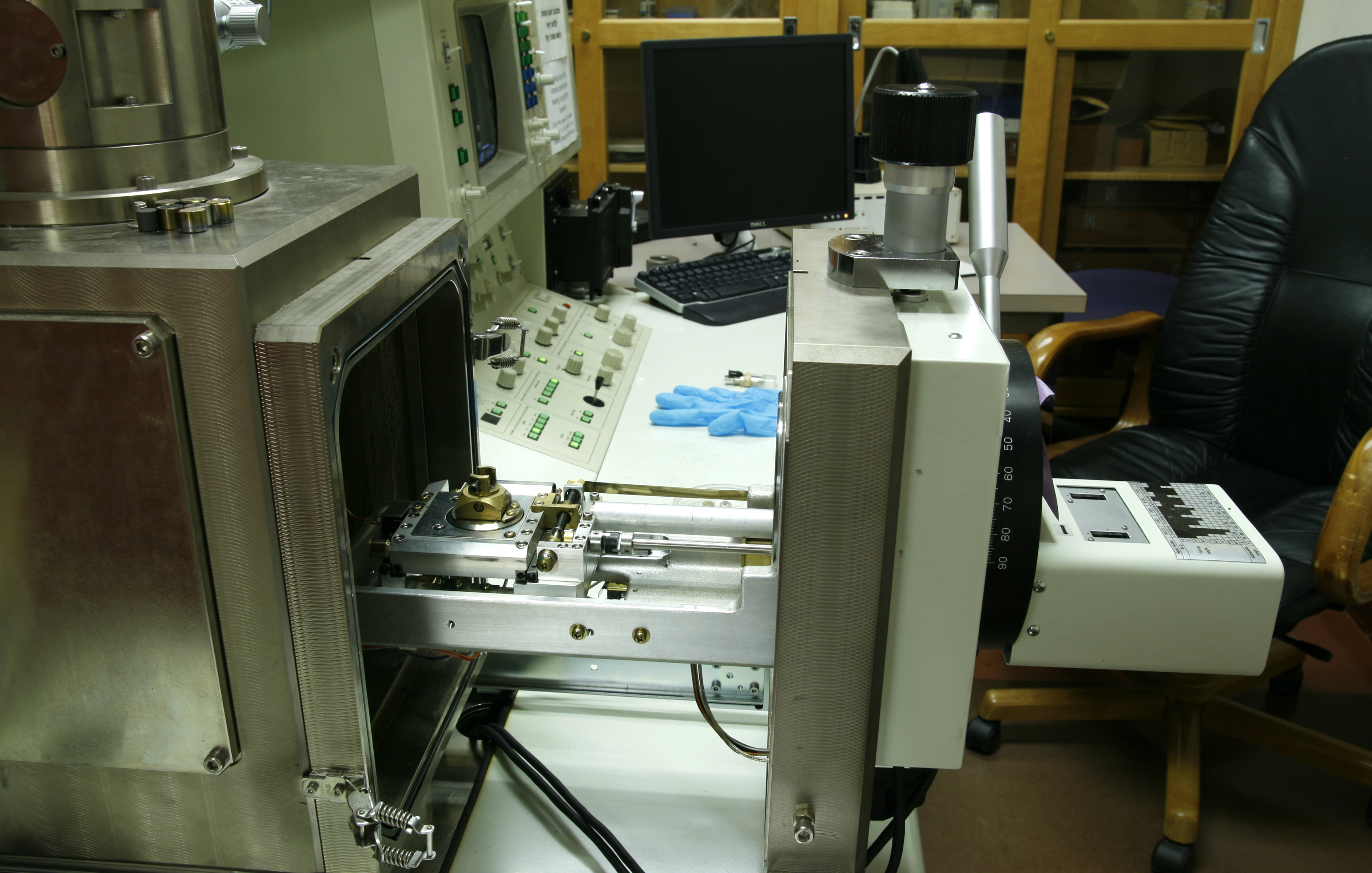
The actual business of the SEM takes place inside of a chamber that houses the various imaging detectors and positioning mechanisms. This chamber is also drawn down to a "hard vacuum" to remove any O2 that would cause the tungsten filament to burn out. The tungsten filament is the source of the electron beam that creates the imaging electrons, thus "Electron" Microscope. The positioning mechanisms are machined in such a way as to accommodate minute angular and rotational movements.
 |
Any organic material not plated with gold will interfere with the imaging process.
This necessitates that handling of the samples must be done while gloved.
Here are his images of A. sporocarpia.
-----------------------------------------------------------------------------------------------------
This is the first batch of images Billy captured with the SEM. The context of the sporocarp (hyphae binding grains of sand) can be seen quite clearly.
 |
 |
 |
The image on the right shows some interesting hyphal detail.
 |
 |
 |
 |
 |
Here is Billy's second batch of images, focusing on the spores.
 |
 |
In these two images can be seen what appear to be empty, deflated spore sacs,
presumably remaining after the mature spores have popped out and dispersed.
 |
 |
 |
 |
 |
 |
 |
 |
Way cool stuff, Mr. Stone. Thank you for sharing! (These images are his ©2011, btw.)
Back to Down and Dirty.
Back to Great Balls O' Dirt.