Here are some photomicrographs by Dr. Chris Walker.
Dr. Walker's comments are included, and for the scales, each small division is equal to 10 Ám (micrometres), each large division is 50 Ám (1/20 mm).
You can click on an image to see a larger version, which may take a minute or two to load.
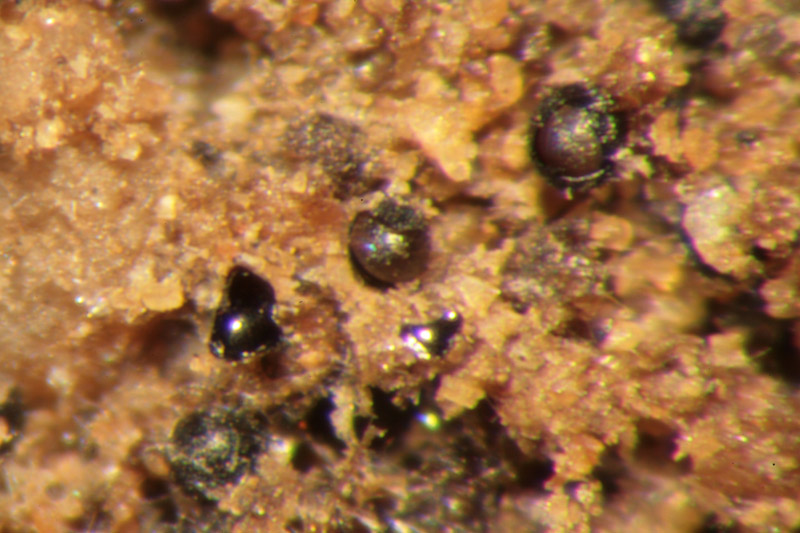
This picture is of a cut surface, showing the spores scattered through the substrate, which consists of sand particles bound together by coarse mycelium. In this image, some of the spores have cracked open like an egg due to the cutting process (with a razor blade).
|
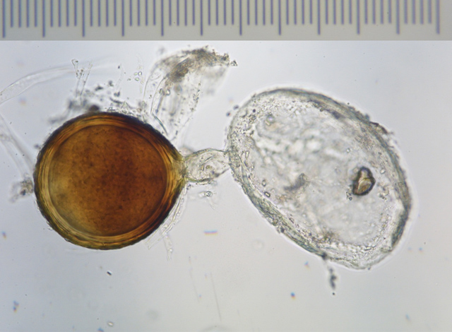
Here is an image of a bleached spore (I have to bleach them to see their structure and developmental characteristics).
 |
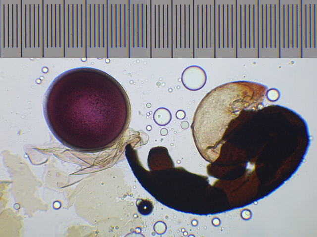
This photo shows the reaction of the inner 'spore' to Melzer's reagent. This is used in the taxonomy of the group, though without too much knowledge of why the reaction takes place (wall polysaccharides). Yours has a nice purple reaction that is found in quite a few things in the order, but I have to say I am now convinced it is not an Acaulospora and will have to be moved to a different (maybe new) genus. Much more work needed before this happens!!!
 |
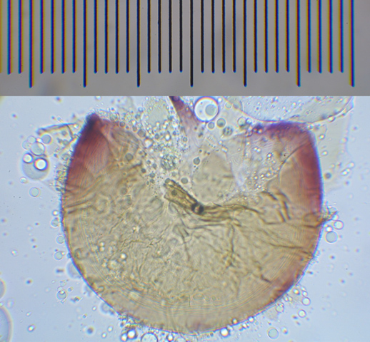
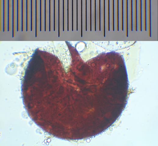
This is the middle of a spore popped out and then put in polyvinyl alcohol lacto-glycerol with Melzer's reagent and ruthlessly squashed. You can just see the reaction beginning to take place at the point of fracture in the first image, and the completed reaction in the second.
 |
 |
Thank you, Dr. Walker, for sharing these amazing images with us.
Back to Great Balls O' Dirt.